2,700-year-old marijuana found in Chinese tomb
TheStar.com - sciencetech - 2,700-year-old marijuana found in Chinese tomb
Stash seems to have been intended for buried shaman to use in the afterlife
November 27, 2008
Dean Beeby
THE CANADIAN PRESS
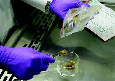
OTTAWA – Researchers say they have located the world's oldest stash of marijuana, in a tomb in a remote part of China.
The cache of cannabis is about 2,700 years old and was clearly ``cultivated for psychoactive purposes," rather than as fibre for clothing or as food, says a research paper in the Journal of Experimental Botany.
The 789 grams of dried cannabis was buried alongside a light-haired, blue-eyed Caucasian man, likely a shaman of the Gushi culture, near Turpan in northwestern China.
The extremely dry conditions and alkaline soil acted as preservatives, allowing a team of scientists to carefully analyze the stash, which still looked green though it had lost its distinctive odour.
"To our knowledge, these investigations provide the oldest documentation of cannabis as a pharmacologically active agent," says the newly published paper, whose lead author was American neurologist Dr. Ethan B. Russo.
Remnants of cannabis have been found in ancient Egypt and other sites, and the substance has been referred to by authors such as the Greek historian Herodotus. But the tomb stash is the oldest so far that could be thoroughly tested for its properties.
The 18 researchers, most of them based in China, subjected the cannabis to a battery of tests, including carbon dating and genetic analysis. Scientists also tried to germinate 100 of the seeds found in the cache, without success.
The marijuana was found to have a relatively high content of THC, the main active ingredient in cannabis, but the sample was too old to determine a precise percentage.
Researchers also could not determine whether the cannabis was smoked or ingested, as there were no pipes or other clues in the tomb of the shaman, who was about 45 years old.
The large cache was contained in a leather basket and in a wooden bowl, and was likely meant to be used by the shaman in the afterlife.
"This materially is unequivocally cannabis, and no material has previously had this degree of analysis possible," Russo said in an interview from Missoula, Mont.
"It was common practice in burials to provide materials needed for the afterlife. No hemp or seeds were provided for fabric or food. Rather, cannabis as medicine or for visionary purposes was supplied."
The tomb also contained bridles, archery equipment and a harp, confirming the man's high social standing.
Russo is a full-time consultant with GW Pharmaceuticals, which makes Sativex, a cannabis-based medicine approved in Canada for pain linked to multiple sclerosis and cancer.
The company operates a cannabis-testing laboratory at a secret location in southern England to monitor crop quality for producing Sativex, and allowed Russo use of the facility for tests on 11 grams of the tomb cannabis.
Researchers needed about 10 months to cut red tape barring the transfer of the cannabis to England from China, Russo said.
The inter-disciplinary study was published this week by the British-based botany journal, which uses independent reviewers to ensure the accuracy and objectivity of all submitted papers.
The substance has been found in two of the 500 Gushi tombs excavated so far in northwestern China, indicating that cannabis was either restricted for use by a few individuals or was administered as a medicine to others through shamans, Russo said.
"It certainly does indicate that cannabis has been used by man for a variety of purposes for thousands of years."
Russo, who had a neurology practice for 20 years, has previously published studies examining the history of cannabis.
"I hope we can avoid some of the political liabilities of the issue," he said, referring to his latest paper.
The region of China where the tomb is located, Xinjiang, is considered an original source of many cannabis strains worldwide.
Moderators: kylervk, Joe, Hank Fist, inx515xhell
-
Big Fat Retard

- Jizzmopper
- Posts: 2999
- Joined: Sat Jul 29, 2006 2:57 pm
- Location: 16th & Jefferson
- Contact:
Dude, where's my stash?
I poop on Petland!

Re: Dude, where's my stash?
Joe was telling me about this on Friday. This story was so ridiculous I almost didn't believe him.
Joey Chaos wrote:Shane's gonna find out the hard way.
-
inx515xhell

- 420
- Posts: 4668
- Joined: Sun Oct 17, 2004 12:34 pm
- Location: denver
- Contact:
Re: Dude, where's my stash?
dibs on this for song material.
FOR REAL.
FOR REAL.
Re: Dude, where's my stash?
Apparently i have to do the real work around here:
Journal of Experimental Botany, Vol. 59, No. 15, pp. 4171–4182, 2008
doi:10.1093/jxb/ern260
This paper is available online free of all access charges (see http://jxb.oxfordjournals.org/open_access.html for further details)
RESEARCH PAPER
Phytochemical and genetic analyses of ancient cannabis
from Central Asia
Ethan B. Russo1,2,3,*, Hong-En Jiang4,5, Xiao Li5, Alan Sutton2, Andrea Carboni6, Francesca del Bianco6,
Giuseppe Mandolino6, David J. Potter2, You-Xing Zhao7, Subir Bera8, Yong-Bing Zhang5, En-Guo Lu¨ 9, David
K. Ferguson10, Francis Hueber11, Liang-Cheng Zhao12, Chang-Jiang Liu4, Yu-Fei Wang4 and Cheng-Sen Li5,13,*
1 Visiting Professor, Institute of Botany, Chinese Academy of Sciences, erusso@gwpharm.com
2 GW Pharmaceuticals, Porton Down Science Park, Salisbury, Wiltshire SP4 OJQ, UK
3 Faculty Affiliate, Department of Pharmaceutical Sciences, University of Montana, Missoula, MT, USA
4 Laboratory of Systematic and Evolutionary Botany, Institute of Botany, Chinese Academy of Sciences, Beijing
100093, China
5 Bureau of Cultural Relics of Turpan Prefecture, Turpan 838000, Xinjiang, China
6 CRA-Centro di Recerca per le Colture Industriali, via di Corticella 133, 40128, Bologna, Italy
7 State Key Laboratory of Phytochemistry and Plant Resources in West China, Kunming Institute of Botany, Chinese
Academy of Sciences, Kunming 650204, China
8 Department of Botany, University of Calcutta, Kolkata 700019, India
9 Xinjiang Institute of Archaeology, 4-5 South Beijing Road, U¨ru¨mqi, Xinjiang 830011, China
10 Institute of Palaeontology, University of Vienna, Althanstrasse 14, A-1090 Vienna, Austria
11 Department of Paleobiology, Smithsonian Institutions, Washington, DC 20560-0121, USA
12 College of Biological Science and Biotechnology, Beijing Forestry University, Beijing 100083, China
13 Beijing Museum of Natural History, Beijing 100050, China
Received 7 August 2008; Revised 24 September 2008; Accepted 25 September 2008
Abstract
The Yanghai Tombs near Turpan, Xinjiang-Uighur
Autonomous Region, China have recently been excavated
to reveal the 2700-year-old grave of a Caucasoid
shaman whose accoutrements included a large cache
of cannabis, superbly preserved by climatic and burial
conditions. A multidisciplinary international team demonstrated
through botanical examination, phytochemical
investigation, and genetic deoxyribonucleic acid
analysis by polymerase chain reaction that this material
contained tetrahydrocannabinol, the psychoactive
component of cannabis, its oxidative degradation
product, cannabinol, other metabolites, and its synthetic
enzyme, tetrahydrocannabinolic acid synthase,
as well as a novel genetic variant with two single
nucleotide polymorphisms. The cannabis was presumably
employed by this culture as a medicinal or
psychoactive agent, or an aid to divination. To our
knowledge, these investigations provide the oldest
documentation of cannabis as a pharmacologically
active agent, and contribute to the medical and
archaeological record of this pre-Silk Road culture.
Key words: Archaeology, botany, cannabis, cannabinoids,
archaeobotany, ethnopharmacology, genetics, medical
history, phytochemistry.
Introduction
Uighur farmers cultivating the land at the base of the Huoyan
Shan (‘Flaming Mountains’) in the Gobi Desert near Turpan,
Xinjiang-Uighur Autonomous Region, China some 20 years
ago uncovered a vast ancient cemetery (54 000 m2) that
seemingly corresponds to the nearby Aidinghu, Alagou, and
* To whom correspondence should be addressed: E-mail: lics@ibcas.ac.cn; erusso@gwpharm.com
ª 2008 The Author(s).
This is an Open Access article distributed under the terms of the Creative Commons Attribution Non-Commercial License (http://creativecommons.org/licenses/by-nc/2.0/uk/) which
permits unrestricted non-commercial use, distribution, and reproduction in any medium, provided the original work is properly cited.
Subeixi excavations (Ma and Wang, 1994; Chen and
Hiebert, 1995; Davis-Kimball, 1998; Kamberi, 1998; An,
2008) (see Supplementary Fig. S1 at JXB online) attributed
to the Gushı culture (later rendered Ju¨shi, or Cheshi)
(Academia Turfanica, 2006). The first written reports
concerning this clan, drafted about 2000 years BP (before
present) in the Chinese historical record, Hou Hanshu,
described nomadic light-haired blue-eyed Caucasians speaking
an Indo-European language (probably a form of
Tocharian, an extinct Indo-European tongue related to
Celtic, Italic, and Anatolic (Ma and Sun, 1994). The Gushı
tended horses and grazing animals, farmed the land
and were accomplished archers (Mallory and Mair, 2000).
The site is centrally located in the Eurasian landmass
(Fig. 1A, B), 2500 km from any ocean and located in the
Ayding Lake basin, the second lowest spot on Earth after
the Dead Sea (Fig. 1A, B). Formal excavations completed
in 2003 revealed some 2500 tombs dating from 3200–2000
years BP (Xinjiang Institute of Cultural Relics and Archaeology,
2004). Other evidence from chipped stone tools and
other items indicate a possible human presence in the area
for some 10 000–40 000 years (Kamberi, 1998; Academia
Turfanica, 2006). Due to a combination of deep graves (2 m
or more), an extremely arid climate (16 mm annual rainfall),
and alkaline soil conditions (pH 8.6–9.1 (Pan, 1996), the
remarkable preservation of the human remains resulted in
the mummification of many bodies without a need for
chemical methods. Numerous artefacts from the tombs
included equestrian equipment and numerous Western
Asian crops such as Capparis spinosa L. (capers) (Jiang
et al., 2007), Triticum spp. (wheat), Hordeum spp. (naked
barley), and Vitis vinifera L. (grapevines) (Jiang, 2008),
often centuries before their first descriptions in Eastern
China (Puett, 1998).
One tomb, M90 (GPS coordinates: 42 48.395# N, 89
38.958# E; elevation, 58 m) (see Supplementary Fig. S2A, B
at JXB online), contained the skeletal remains of a male
of high social status of an estimated age of 45 years, whose
accoutrements included bridles, archery equipment, a kongou
harp, and other materials supporting his identity as
a shaman (see Supplementary Figs S3A, B, 4A–C at JXB
online). His burial as a disarticulated skeleton, as opposed
to a mummified body as more frequently was found,
suggested that he probably died in the highlands of the Tian
Shan (‘Heavenly Mountains,’ or Ta¨ngri Tagh in Uighur)
(Fig. 1), and his bones were later interred at Yanghai, as
Fig. 1. Area maps. (A) Map of Turpan, Xinjiang, China and its location in Central Asia. (B) Map of Yanghai Tombs site and surrounding area
(adapted from Xinjiang Institute of Cultural Relics and Archaeology, 2004).
4172 Russo et al.
nearby tombs contained large timbers of Picea (spruce)
spp. that grow at 3000 m elevation. Modern Uighur
pastoralists follow a similar annual migratory path to
summer grazing lands some 60–80 km distant from the
tombs. Near the head and foot of the shaman’s bier lay
a large leather basket and wooden bowl (see Supplementary
Fig. S5A, B at JXB online) filled with 789 g of vegetative
matter, initially thought to be Coriandrum sativum L.
(coriander), but which, after meticulous botanical examination,
proved to be Cannabis sativa L. (Jiang et al., 2006).
An initial radiocarbon date of 2500 years BP has subsequently
been corrected to a calibrated figure of 2700
years BP based on additional analyses of equestrian gear and
correlation to tree ring data (dendrochronology) in China.
While an earlier publication (Jiang et al., 2006) emphasized
morphological features in identifying the cannabis, the
current study used additional botanical, phytochemical, and
genetic investigations to demonstrate that this cannabis was
psychoactive and probably cultivated for medicinal or
divinatory purposes. Great care was taken to prevent
contamination of the sample throughout the analyses.
Materials and methods
Photomicrography methods
Upon courier delivery from China, a polythene bag containing 11 g
of ancient cannabis was sterilized with ethanol, handled with
laboratory gloves in a laminar-flow hood, and transferred with a clean
metal spatula (Fig. 2A). Two levels of light microscopy were used in
this study. For the observations on the achenes (Fig. 2D), a low
power Brunel MX3 microscope (Chippenham, Wiltshire, UK) was
used and a 33 objective utilized in conjunction with an Olympus
SP350 8 megapixel camera, stereo insert 30 mm lens tube, and
Photonic PL2000 – double arm cold light source. Greater magnification
was required for more detailed observations of trichomes
(Fig. 2B, C): a high power stereo light microscope with a Trinocular
Head for camera attachment (STE UK, Sittingbourne, Kent, UK)
with an eye piece graticule for specimen size measurement fitted
with 34, 310, and 340 objectives. The camera’s 33 optical zoom
capability provided additional magnification.
The observations on the seed were made on unmounted specimens.
For these, small pieces of plant tissue were placed directly onto
the low-power microscope plate. When using the high power
microscope, samples were dry mounted on a glass slide. To achieve
views where large proportions of the material were simultaneously in
focus, flat samples specimens (as shown) gave the greatest success.
On the low power microscope the seed sample was illuminated
with incident light, using a Photonic PL2000 – double arm ‘cold
light source’ (Fig. 2D). Some samples, when placed on the high
power microscope, were also illuminated using the cold light
source. Others were illuminated from below. When viewing
samples mounted beneath a cover slip, it is common to set up
a microscope using the Ko¨hler illumination method (Delly, 1988).
This ensured that light from the condenser lens was focused
correctly on the microscope slide. For uncovered specimens, the
condenser height and aperture were adjusted while viewing
the subject until optimum resolution was achieved. In all cases, the
specimens were measured using a graticule within the eyepiece.
To enable photographs to be taken through the low power
microscope, one eyepiece was replaced with a compatible 30 mm
Fig. 2. Photomicrographs of ancient cannabis. (A) Photograph of the whole cannabis sample being transferred in laminar flow hood. (B)
Photomicrograph of leaf fragment at low power displaying non-glandular and amber sessile glandular trichomes. Note retention of chlorophyll and
green colour, scale bar¼100 lm. (C) Higher power photomicrograph of a single sessile glandular trichome. At least 4 of its 8 secretory cells are
clearly visible on the right, and the scar of attachment to the stype cells in the centre, scale bar¼25 lm. (D) Low power photomicrograph of
a cannabis achene (‘seed’) including the base with a non-concave scar of attachment visible, scale bar¼1 mm.
Ancient cannabis 4173
lens tube to which single lens reflex or digital cameras would be
attached. As in ordinary photography, the depth of field is
considered to be the distance from the nearest object plain to the
farthest object plain that is in focus. When objects are a long
distance from the camera lens the depth of field is large. However,
depth decreases as the image comes closer to the lens. When taking
photomicrographs, depth of field is measured in microns (Delly,
1988). To maximize the chance of finding substantial areas of tissue
simultaneously in focus within this narrow depth of field, multiple
samples were laid as flat as possible onto glass slides. In all cases,
photomicrographs were taken on a solid bench and the shutter
activated remotely to reduce manually-induced camera-shake.
In no instance was any image modification technique used in
these photographs.
Phytochemistry methods
Approximately 2 g of the dried plant material was extracted with
200 ml methanol:chloroform (9:1 v/v) by sonication at room
temperature (21 C), the standard extractive technique for this
laboratory (GW Pharmaceuticals), a method that recruits >95% of
phytocannabinoid content. The solvent layer was then transferred
through a paper filter into a rotary evaporator flask. The flask was
evaporated to dryness at 40 C, under reduced pressure, prior to
resuspension in 4 ml of methanol:dichloromethane (3:1 v/v). This
sample was transferred to two autosampler vials to be analysed by
GC-FID-MS and HPLC-UV. At all stages, the clean glassware was
extracted with the same solvents to ensure that none of the observed
peaks would be a result of contamination. GC-FID-MS analyses
were performed on a HP6890 gas chromatograph, coupled to a 5975
inert mass spectrometer. The system was controlled with Agilent
MSD chemstation D.03.00.611. The GC was fitted with a Zebron
fused silica capillary column (30 m30.32 mm inner diameter)
coated with ZB-5 at a film thickness of 0.25 lm (Phenomenex). The
oven temperature was programmed from 70 C to 305 C at a rate of
5 C min1. The injector port and the transfer line were maintained
at 275 C and 300 C, respectively. Helium was used as the carrier
gas at a pressure of 55 kPa. The injection split ratio was 5:1. HPLC
profiles were obtained using an Agilent 1100 series HPLC system
controlled by Chemstation version A09.03 software. Cannabinoid
profiles were generated using a C18 (15034.6 mm, 5 lm) analytical
column fitted with a C18 (1034.6 mm, 5 lm) guard column. The
mobile phase consisted of acetonitrile, 0.25% w/v acetic acid and
methanol at a flow rate of 1.0 ml min1 and the column was kept at
35 C. The UV profiles were recorded at 220 nm.
Genetic methods
DNA was extracted from pulverized dried leaves, from two seeds
probably belonging to Cannabis spp., and from three seeds probably
from other unidentified species. The DNeasy Plant Mini Kit (Qiagen)
was used, according to the Qiagen protocol, but with some
modification to increase the final DNA amount and to avoid external
and artificial contamination. For this reason, pre-PCR and post-PCR
operations were physically separated and carried out in different
environments. Ancient DNA extraction and other pre-PCR works
were performed under a UV-filtered ventilation system and a positive
pressure airflow. Filtered pipette tips and sterile tubes and plastics
were always used; gloves, masks, and laboratory coats were always
worn. The quality of DNA obtained was estimated by A260/A280
absorbance ratio. In order to obtain the highest possible fidelity
during PCR synthesis, PCR reactions were performed using the Pwo
Master ready-to-use proofreading master mix (Roche Applied
Science) according to their protocol. The primers designed to test
DNA integrity and suitability for PCR analysis and species
identification were from the ITS region of nuclear ribosomal DNA
(Blattner, 1999), and from a non-coding region of chloroplast DNA
(Taberlet et al., 1991). The reaction mixtures were subjected firstly to
an initial heat denaturation at 94 C for 3 min; then, they were
subjected to 35 cycles of heat denaturation at 94 C for 30 s, 1 min of
primer annealing at 55 C for the ITS region, and 50 C for cpDNA,
and DNA extension at 72 C for 40 s. Finally, the samples were
maintained at 72 C for 5 min for the final extension. PCR reactions
were performed in an MJ Research PTC-100 thermal cycler (MJ
Research, USA). The amplification products were separated by
electrophoresis in a 1.5% agarose gel. The bands were excised and
purified with the MinElute Gel Extraction Kit (Qiagen). PCR-purified
products were quantified and directly forward- and reversesequenced,
using the GenomeLab Dye Terminator Cycle Sequencing
with a Quick Start Kit on a CEQ8000 Genetic analyser (Beckman
Coulter). Primer sequences were identified and removed manually,
and database searches were performed with the BLASTN algorithm
(Altschul et al., 1990). The sequences results proved that the
pulverized dried tissue was from Cannabis sativa L., despite our
observation in the mixed sample of some small seeds of different
species, removed before the DNA extraction; no differences were
observed between the sequences obtained and those deposited at the
NCBI gene-bank (for THCA-and CBDA-synthases, GeneBank
accession numbers E55108/GI 18529739 and E33091/GI 18623981).
By contrast, no amplification was obtained from DNA extracted from
seeds of both cannabis and the other, unidentified species. The allelic
status at a single locus, B, known to be the major gene determining
the CBD/THC ratio in cannabis (de Meijer et al., 2003), was
investigated in the ancient material. The primer pairs described (de
Meijer et al., 2003) are not sufficiently associated with the chemotype
(Pacifico et al., 2006), and the sequence-based primers described
therein (Pacifico et al., 2006) failed to yield any amplification,
probably due to the limited integrity of DNA from ancient cannabis
tissues, which did not sustain the amplification of a 1100 Da DNA
fragment. Therefore, three different primer pairs (Fw1503Rev328,
Fw1663Rev318, and Fw1543Rev318) were used. These primers
were designed on two conserved small regions of a zone varying
between the known sequences of THC and CBD alleles. When tested
on fresh cannabis tissues, these primers were demonstrated to be able
to amplify both alleles (PCR and sequences data not shown). Using
different primer pair combinations, the risk of a no-match or
a mismatch because of possible mutations in the 3# end of primer
region was overcome. The primer sequences are listed in Supplementary
Fig. S8 at JXB online. All reaction mixtures were subjected first
to heat denaturation at 94 C for 3 min and then to 35 cycles
consisting of heat denaturation at 94 C for 15 s, primer annealing at
54 C for 30 s, and DNA extension at 72 C for 1 min. Finally, the
samples were maintained at 72 C for 5 min for the final extension of
DNA. PCR products were separated by electrophoresis in a 1.5%
agarose gel. The bands were excised and purified with MinElute Gel
Extraction Kit (Qiagen). PCR-purified products were quantified and
directly sequenced in forward and reverse, using the GenomeLab
Dye Terminator Cycle Sequencing with Quick Start Kit on
a CEQ8000 Genetic analyser (Beckman Coulter).
Results
Microscopic botanical analysis
Gross examination of the 11 g sample of cannabis provided
by the Chinese Academy of Sciences revealed loose dry
vegetative material (Fig. 2A). The impression that the
vegetative material had been lightly pounded was supported
by examination of the wooden bowl, whose internal
surface was worn smooth, apparently from use as a mortar
(see Supplementary Fig. S5B at JXB online). The cannabis
4174 Russo et al.
retained a surprisingly green colour in its leafy parts and
displayed visible glandular trichomes (Fig. 2B), the
phytochemical factory of the plant and site of manufacture
of cannabinoids and terpenoids (Potter, 2004; McPartland
and Russo, 2001; Kim and Mahlberg, 2003). However, the
ancient sample lacked the typical cannabis odour. Microscopic
examination confirmed the presence of intact sessile
trichomes with an amber tint (Fig. 2B), while higher
resolution documented the retention of visible secretory
cells within the trichomes (Fig. 2C). Achenes (‘seeds’)
averaged 2.2–3.6 mm in length (Jiang et al., 2006), were
light in colour with some striations, but demonstrated
rough, non-concave fruit attachment (Fig. 2D), all traits of
domestication (Schlumbaum et al., 2008) associated with
cultivated cannabis strains (Vavilov, 1926). In contrast,
achenes of wild strains are typically smaller and darker
with concave attachment zones that favour shattering and
easy spread (Vavilov, 1926). Germination was attempted
with 100 achenes in compost, but no emergence was
observed after 21 d.
Phytochemical analysis
Phytochemical and genetic teams were initially blinded to
one another’s results. The extraction of 2 g of plant
material produced 67.9 mg of solids after the removal of
solvents. Using high performance liquid chromatography
(HPLC), the largest cannabinoid peak was cannabinol
(CBN) at 7.4 min, but concentration levels were very low,
averaging 0.007% w/w. CBN is an oxidative breakdown
product THC, generated non-enzymatically, with increasing
age (Brenneisen, 2007). There were also peaks
corresponding to expected retention times for cannabidiol
(CBD) at 4.9 min and cannabichromene (CBC) at 12 min
(Fig. 3). Both are phytocannabinoids resulting from
alternative enzymatic pathways than that yielding THC
(de Meijer et al., 2003). There were very few peaks in the
first 20 min of the gas chromatogram where mono- and
sesquiterpenes elute (Fig. 4). This lack of terpenoid
volatiles supports the physical observation that the plant
material lacked the herbal smell traditionally associated
with cannabis (McPartland and Russo, 2001). Shown in
Fig. 5A–C, (and in Supplementary Fig. S7A, B at JXB
online) are breakdowns of sub-regions of the gas
chromatogram. The major peaks in the 13–30.5 min
region are free fatty acids (see Supplementary Fig.S7A at
JXB online). The largest peak identified as palmitic acid
was the most abundant in the sample. Methyl and propyl
cannabinoids eluted in the 27–30 min region and the
Fig. 3. Complete high performance liquid chromatography (HPLC) of ancient cannabis.
Fig. 4. Complete gas chromatography-flame ionization detection (GC-FID) of ancient cannabis.
Ancient cannabis 4175
peaks marked as 286 Da and 302 Da all had MS spectra
consistent with propyl cannabinoids. There were two
phthalate peaks at approximately 23.5 min (believed to
have originated from the polythene bags in which the
samples were supplied). A number of phytocannabinoids
were identified in the 30–34 min region (Fig. 5A)
including cannabidiol (CBD), cannabichromene (CBC),
cannabicyclol (CBL, a heat-generated artefact of CBC
Fig. 5. Gas chromatography of ancient cannabis subsections. (A) GC of the 30–34 min region demonstrates several phytocannabinoids: cannabidiol
(CBD), cannabichromene (CBC), cannabicyclol (CBL), and cannabinavarin (CBNV). (B) GC of the 34–36.3 min region displays the highest peak,
cannabinol (CBN), the direct non-enzymatic oxidative metabolite of THC, with possible cannabielsoin (CBE) at 34.2 min. (C) GC of the 36.3–40.5
min region displays cannabitriol (CBO) a THC degradant, and CBN variants (see text).
4176 Russo et al.
(Brenneisen, 2007), and cannabinavarin (CBNV, a propyl
analogue of CBN). In the 34–36.3 min region (Fig. 5B),
apart from cannabinol (CBN), the largest individual
phytocannabinoid component, there were at least four
peaks of 330 Da with cannabielsoin (CBE, an artefact
derived from CBD (Brenneisen, 2007) a likely identification
of the peak at 34.2 min. In the 36.3–40.5 min region
(Fig. 5C), the known THC degradant cannabitriol (CBO)
(Brenneisen, 2007) was seen, as well as a series of peaks
with spectral similarities to CBN, three of which are
tentatively identified by the NIST database as either
hydroxyl- or oxo-CBN. The last region (42–50 min; see
Supplementary Fig. S7B at JXB online), contained
phytosterols and triterpene alcohols with beta-sitosterol
the most abundant compound.
Mass spectra (MS) of selected phytocannabinoids
corresponding to the above are displayed (Fig. 6). Values
are all in agreement with those in NIST and GW
Pharmaceutical databases.
There was a very small peak detected at the correct
retention time in the sample for THC, but the spectra
could not confirm its identity.
Genetic analysis
Because of the unique degree of preservation of the
cannabis, a genetic analysis was undertaken. The two
ancient DNA sequences determined were labelled China
F and China F(h). Alignment of these paleo-sequences
(excluding the primers’ region, in red in Fig. 7A) with the
presently available databases demonstrated:
(i) China F (Fig. 7A) is identical 134/134 nucleotide
agreement to other deposited sequences: AB212841,
AB212839, AB212836, AB212833, AB212830, all belonging
to tetrahydrocannabinolic acid synthases (THCAsynthases),
a species-specific genetic region (Schlumbaum
et al., 2008) from Cannabis sativa L.
(ii) China F(h) (Fig. 7A) is a new variant, not previously
present in the genetic databases (submitted to NCBI,
GenBank accession number EU839988), showing a maximum
identity of 132/134 nucleotides with the abovementioned
sequences and with China F.
Utilizing BLASTX, i.e. performing searches through
amino acid translation, it was again shown that the China
F(h) amino acid sequence is not registered in the database,
and this is an obvious but necessary confirmation of the
originality of this variant of the THCA synthase allele.
These results also prove that both sequences encode for
THCA synthase, the biosynthetic enzyme for THCA that
decarboxylates via heat or ageing to yield psychoactive
THC (Russo, 2007). Direct comparison of the two ancient
sequences, identified the nature of the small differences
observed: the samples have two ‘mutations’ (highlighted
in yellow in Fig. 7A), which can be considered transversions:
from guanine to cytosine, and from cytosine to
adenine. The first of these two nucleotide substitutions is
synonymous, i.e. it does not change the amino acid
sequence, while the second one is a non-synonymous
substitution, leading to a serine-threonine exchange
(highlighted in light blue in Fig. 7B) in the encoded
amino acid sequence; these two amino acids, however,
have similar physico-chemical properties. No CBDA
synthase, the biosynthetic enzyme for CBD (de Meijer
et al., 2003), was identified in the sample.
Discussion
The results presented collectively point to the most
probable conclusion which is that the Gushı culture
cultivated cannabis for pharmaceutical, psychoactive or
divinatory purposes. In examining the botanical evidence
from this ‘old and cold’ site with its unique degree of
preservation, the cannabis consisted of a processed
(pounded) sample whose seed size, colour, and morphology,
at least according to principles of Vavilov (Vavilov,
1926), suggest that it was cultivated rather than merely
gathered from wild plants. The considerable amount of
cannabis present (789 g) without any large stalks or
branches would logically imply a pooled collection rather
than one from a single plant. Importantly, no obvious
male cannabis plant parts (e.g. staminate flowers, not
infrequently observed in Indian herbal cannabis, or bhang
(Russo, 2007) were evident, implying their exclusion or
possible removal by human intervention, as these are
pharmacologically less psychoactive.
The HPLC, GC, and MS analyses confirm the identity
of the supplied plant sample as Cannabis sativa L. The
predominance of CBN indicates that the original plants
contained D9-tetrahydrocannabinol (THC) as the major
phytocannabinoid constituent. The presence of CBO and
numerous CBN-related substance peaks further supports
this view. CBD and CBC, together with their known
thermo-oxidative degradation products CBE and CBL
(Brenneisen, 2007), are present, but the GC analysis
would appear to indicate that, in both cases, CBC and
CBL are represented in greater quantities, as expected in
a high-THC cannabis strain wherein CBD is only a minor
component. In addition, there is a peak for CBNV which
confirms that the plant also contained D9-tetrahydrocannabivarin
(THCV), a propyl phytocannabinoid. All of these
observations are consistent with strains of cannabis with
a high THC content and in an alternative taxonomy
suggests it should be assigned to Cannabis indica
Lamarck (Hillig and Mahlberg, 2004).
While chromatography elution times may vary with
temperature, column type, and other factors, confirmation
was evident with corroboratory mass spectra values that
Ancient cannabis 4177
were identical to those seen daily in assays performed on
fresh cannabis extracts in this laboratory.
The presence of so many recognized cannabinoid
degradants is consistent with very old cannabis samples.
The very low concentration levels measured in the HPLC
analysis may indicate that the sample provided contained
significantly more leaf and twig material than flower
material, rather than being evidence in itself that the sample
was of low potency originally. This plant material is
therefore conclusively cannabis derived from a population
of plants within which THC was the dominant cannabinoid.
By contrast, a sample taken from a mix of wild-type
Cannabis sativa would customarily harbour a more equal
mixture of THC and CBD (de Meijer et al., 2003). It would
appear, therefore, that humans selected the material from
plants on the basis of their higher than average THC
content. To elaborate, a chemotaxonomy of cannabis
previously outlined indicates three types (Small and
Beckstead, 1973): chemotype I (drug) strains with high-
THC:CBD ratios, chemotype II low-THC, higher-CBD
Fig. 6. Mass spectra of ancient cannabis. Subsections demonstrate the phytocannabinoids cannabinol (CBN), cannabidiol (CBD), cannabicyclol
(CBL), cannabinolivarin (CBNV), cannabichromene (CBC), cannabielsoin (CBE), 1#-oxcannabinol, and 1#-hydroxycannabinol.
4178 Russo et al.
(fibre) strains, and chemotype II with more equal ratios.
THC and CBD production are mediated by co-dominant
alleles BT and BD, respectively (de Meijer et al., 2003). By
comparison, pooled samples from cannabis fields in
Morocco and Afghanistan will normally produce 25%
high-THC plants, 25% high-CBD plants, and 50% with
lower, mixed titres, combining to yield roughly equivalent
amounts of the two phytocannabinoids (Russo, 2007),
a pattern not observed in our specimen.
Isotopic analysis of cellulose from this cannabis sample
might conceivably be used in comparison with other
samples in an attempt to establish its geographic origin.
While multi-purpose cannabis plants used simultaneously
for food (seed), fibre (stalks), and pharmaceutical
uses (flowering tops) have been recently reported from
Darchula in far western Nepal (Clarke, 2007), more
customarily, a given plant is best suited toward a single
purpose. Of additional key importance is the absence of
hemp artefacts from the Yanghai Tombs. The Gushı
fabricated clothing from wool (see Supplementary Fig.
S6B at JXB online) and ropes from Phragmites (reed) spp.
fibres (see Supplementary Fig. S6C at JXB online).
Whereas hemp textiles have been collected from the
Northern China Yangshao Culture from 6000–7000 years
BP, their appearance in the west was not documented
before 2000 years BP, for example, 1500 years BP in
Kucha, 600 km west of Turpan (Mallory and Mair, 2000).
Previous phytochemical analyses of antique cannabis
preparations have demonstrated THC remnant fingerprints
from 19th century cannabis preparations (Harvey, 1990)
including a 140-year-old sample of Squire’s Extract
(Harvey, 1985). A study in 1992 reported the presence of
D8-THC (previously termed D6-THC) from burned cannabis
that was reportedly inhaled as an aide to childbirth in
a Judean cave 1700 years BP (Zias et al., 1993), supported
by the finding of cannabinoid residues in an adjacent glass
vessel (Zias, 1995). In the Mustang region of Nepal,
mummified human remains of probable Mongolian ancestry
have been dated 2200–2500 years BP in association with
cannabis, probably transported from elsewhere (Kno¨rzer,
2000; Alt et al., 2003), but with insufficient detail to
ascertain its use. Rudenko recovered cannabis seeds,
censers, and hempen clothing in Pazyryk, Siberia from
Scythian kurgans (burial mounds) from 2400–2500 years BP
(Rudenko, 1970; Brooks, 1998), closely matching Herodotus’
descriptions of funeral rites for that culture (Herodotus,
1998). Sarianidi also claimed cannabis use in the Bactria–
Margiana Archaeological Complex (BMAC) (present day
Turkmenistan) (Sarianidi, 1994, 1998), but this interpretation
has been debated (see discussion in Russo, 2007).
Another independent genetic analysis of this material
published subsequent to our analysis (Mukherjee et al.,
2008) confirmed the presence of THCA synthase, but not
the single nucleotide polymorphisms. The authors posited
a European–Siberian origin for the material.
Current genetic data also confirm that the plant material
examined is Cannabis sativa L. according to ITS and
cpDNA analysis. The results also support the hypothesis
Fig. 7. DNA analysis of ancient cannabis. (A) Nucleotide sequences of the wild-type tetrahydrocannabinolic acid synthase, China F, and the mutant
sequence, China F(h), with two single nucleotide polymorphisms highlighted in lower case yellow. (B) Amino acid translation of China F and China
F(h), demonstrating divergence in a change from serine (wild-type) to threonine (mutant), highlighted in blue.
Ancient cannabis 4179
of the existence of at least two THCA-synthase nucleotide
sequences in the ancient plant material examined. One of
these sequences perfectly matches the corresponding
sequence of already-known THCA-synthases deposited in
GenBank, both as gene and protein sequences; the second
sequence is a novel one, with two single nucleotide
polymorphisms (SNPs) encoding for a protein with
presumably very similar characteristics. Whether these
two sequences coexisted in a single cannabis plant or
a strain heterozygous at the B locus, or belong to different
plants, could not be concluded.
THC represents one of the possible phytocannabinoid
end-products manufactured by cannabis plants; THC (or, in
its native form, THCA) is synthesized by a well-characterized
enzyme (THCA- or THC-synthase) from a precursor
(CBG or CBGA) common to most chemotypes that
represents the metabolic ‘switching point’, downstream of
which the variability of the different chemotypes is
concentrated. The agents of such variability found in
cannabis germplasm are exclusively the different synthases,
among which THC(A)-synthase is the only one responsible
for making that specific cannabinoid, THC. Therefore, the
presence of the allelic variant responsible for coding the
THC(A)-synthase may well be considered to be diagnostic,
or at least strongly suggestive of a THC-producing plant.
The fossil cannabis plants found were therefore genetically
equipped to produce THC. How much THC they actually
produced, cannot of course be specified because they
depend on a number of anatomical, environmental, and
nutritional factors that remain unknown.
Numerous questions remain. Current data do not permit
it to be ascertained how the cannabis from the tomb was
administered. If used orally, perhaps it was combined in
some fashion with Capparis spinosa L., as these plants
were found together in a nearby but later tomb at Yanghai
(Jiang et al., 2007). That date for that tomb was initially
reported as 2700 years BP via radiocarbon methods, and
since corrected to 2200–2400 years BP with additional
calibration employing tree ring data. If this cannabis were
smoked or inhaled, no mechanism for so doing has been
excavated in the area. The Gushı could have sifted the
cannabis through fabric after pounding, then fumigated it,
much as described for the alleged cannabis candidate, the
Sumerian A.ZAL.LA, administered medicinally for ‘hand
of ghost’(Thompson, 1923, 1949), since posited as
nocturnal epilepsy (Russo, 2007; Wilson and Reynolds,
1990). While this culture could have arrived from the
earlier BMAC region as ‘oasis hoppers’ (Barber, 1999),
and certain cultural relationships are apparent to the
Scythian culture with respect to cannabis use and
equestrian prowess, those peoples were Iranian speakers
(Mallory and Mair, 2000). In addition, Gushı cultural
affinities and burial practices much more closely resemble
those of the presumed proto-Tocharian speaking, incenseburning
(Kuzmina, 1998) Afanasievo peoples in the
Yenisei Valley to the north (Anthony, 1998, 2007;
Mallory, 1998; Renfrew, 1998; Mallory and Mair, 2000),
whose putative southward migration some authorities
have attributed to ‘global cooling’ c. 4000 years BP (Hsu¨,
1998), and to their proto-Indo-European-speaking Yamnaya
forebears further west, dating to 6000 years BP
(Mallory, 1989; Anthony, 1998; Winter, 1998). Abundant
mysteries remain as to the origins and customs of the
Gushı. Additional answers may accrue from future
archaeological excavations or human genetic analyses that
elucidate relationships with other ancient cultures and
modern peoples of the region. The unique SNPs discovered
in this ancient sample may yet be of critical
importance in tracing the phylogeny and geographic
spread of cannabis and the humans who used it.
The excellent preservation of the cannabis from this tomb
allowed an unprecedented level of modern botanical
investigation through biochemistry and genetics to conclude
that the plant was cultivated for psychoactive
purposes. While cultivation of hemp for fibre has been
documented in Eastern China from a much earlier date
(vide supra Mallory and Mair, 2000), the current findings
represent the most compelling physical evidence to date for
the use of cannabis for its medicinal or mystical attributes.
Supplementary data
Photographs and diagrams of the Yanghai Tombs site,
Tomb M90 contents including fabric and ropes, and
additional chromatographic and genetic analysis primer
sequence information are presented in Supplementary Figs
S1–S8, available online.
Fig. S1. Study site at the Yanghai tombs with Huoyan
Shan mountain range in background (photo EBR).
Fig. S2. Diagrams of the Yanghai Tombs (adapted from
Xinjiang Institute of Cultural Relics and Archaeology,
2004, with permission).
Fig. S3. The shaman’s tomb, M90 [previously published
in Mandarin (Xinjiang Instgitute of Cultural Relics
and Archaeology, 2004), used with permission].
Fig. S4. The shaman’s skull (photos EBR).
Fig. S5. Containers in which cannabis was stored in
tomb [previously published in Mandarin (Xinjiang Institute
of Cultural Relics and Archaeology, 2004] used
with permission.
Fig. S6. Re-excavation of Tomb M90. This was
undertaken to re-examine artefacts, measure GPS coordinates,
and assess environment conditions (photos
EBR).
Fig. S7. Chromatography subsections from phytochemical
analysis.
Fig. S8. Primer sequences employed in the genetic
analysis to amplify THC- and CBD-allele specific fragments
and their sequences (5#/3#).
4180 Russo et al.
Acknowledgements
The authors are grateful to the Chinese Academy of Sciences and
GW Pharmaceuticals for support of the project. Kim Laughton
facilitated communication and logistics between the Chinese
authorities and the British Home Office for exportation and
importation of the ancient cannabis. Daniel Adams, Laura-Jane
Everitt, and Helen Keogh performed phytochemical analytical
preparation, supervised by Peter Gibson. Ying Li provided translation
and logistical support to EBR during field work in Xinjiang.
Gregory Gerdeman is thanked for his helpful review of the article,
as are the anonymous reviewers for their suggestions. No competing
financial interests were operative in this study.
Author contributions: EBR proposed and co-ordinated the current
study, engaged in field work, and wrote the article drafts. HEJ
performed the majority of the background research and was actively
engaged in current investigations. AS performed the phytochemical
analysis and wrote the pertinent methods and results sections. AC,
FDB, and GM performed the genetic analysis and wrote the
pertinent methods and results sections. DJP co-ordinated
the handling of the ancient cannabis in the UK, performed the
microphotography, and wrote the pertinent methods and results.
EGL, XL, DKF, FH, YBZ, YFW, LCZ, and CJL were all engaged
in earlier investigations in relation to this study. YXZ analysed the
phytochemistry of the cannabis sample and SB and his colleagues
analysed the phytochemistry and genetics of the cannabis sample
independently. CSL conceived the concept of studying the
archaeological cannabis samples by multidisciplinary methods, and
organized, co-ordinated and supervised all aspects of the current
study and its performance.
References
Academia Turfanica. 2006. Selected treasures of the Turfan relics.
Turpan, China: Academia Turfanica.
Alt KW, Burger J, Simons A, et al. 2003. Climbing into the past:
first Himalayan mummies discovered in Nepal. Journal of
Archaeological Science 30, 1529–1535.
Altschul SF, Gish W, Miller W, Myers EW, Lipman DJ. 1990.
Basic local alignment search tool. Journal of Moleculr Biology
215, 403–410.
An Z. 2008. Cultural complexes of the Bronze Age in the Tarim
Basin and surrounding areas. In: Mair VH, ed. The Bronze Age
and Early Iron Age peoples of Eastern Central Asia, Vol. I.
Washington, DC: Institute for the Study of Man, 45–62.
Anthony DW. 1998. The opening of the Eurasian steppe at 2000
BCE. In: Mair VH, ed. The Bronze Age and Early Iron Age
peoples of Eastern Central Asia, Vol. I. Washington, DC:
Institute for the Study of Man, 94–113.
Anthony DW. 2007. The horse, the wheel, and language: how
Bronze-Age riders from the Eurasian steppes shaped the modern
world. Princeton, NJ; Oxford: Princeton University Press.
Barber EJW. 1999. The mummies of U¨ ru¨mchi. New York: WW
Norton & Company.
Blattner FR. 1999. Direct amplification of the entire ITS region
from poorly preserved plant material using recombinant PCR.
Biotechniques 27, 1180–1186.
Brenneisen R. 2007. Chemistry and analysis of phytocannabinoids
and other Cannabis constituents. In: Elsohly M, ed. Marijuana
and the cannabinoids. Totowa, NY: Humana Press, 17–49.
Brooks EB. 1998. Textual evidence for 04c Sino-Bactrian contact.
In: Mair VH, ed. The Bronze Age and Early Iron Age peoples of
Eastern Central Asia, Vol. II. Washington, DC: Institute for the
Study of Man, 716–726.
Chen K-T, Hiebert FT. 1995. The late prehistory of Xinjiang in
relation to its neighbors. Journal of World Prehistory 9, 243–300.
Clarke RC. 2007. Traditional Cannabis cultivation in Darchula
District, Nepal: seed, resin and textiles. Journal of Industrial
Hemp 12, 19–42.
Davis-Kimball J. 1998. Tribal interactions between the Early Iron
Age nomads of the southern Ural steppes, Semirechiye, and
Xinjiang. In: Mair VH, ed. The Bronze Age and Early Iron Age
peoples of Eastern Central Asia, Vol. I. Washington, DC:
Institute for the Study of Man, 238–263.
de Meijer EP, Bagatta M, Carboni A, Crucitti P, Moliterni VM,
Ranalli P, Mandolino G. 2003. The inheritance of chemical
phenotype in Cannabis sativa L. Genetics 163, 335–346.
Delly JG. 1988. Photography through the microscope. Rochester,
NY: Eastman Kodak.
Harvey DJ. 1985. Examination of a 140 year old ethanolic extract
of Cannabis: identification of new cannabitriol homologues and
the ethyl homologue of cannabinol. In: Harvey DJ, Paton W,
Nahas GG, eds. Marihuana ‘84. Proceedings of the Oxford
symposium on Cannabis. Oxford, UK: IRL Press, 23–30.
Harvey DJ. 1990. Stability of cannabinoids in dried samples of
cannabis dating from around 1896–1905. Journal of Ethnopharmacology
28, 117–128.
Herodotus. 1998. The histories. Oxford [England]; New York:
Oxford University Press.
Hillig KW, Mahlberg PG. 2004. A chemotaxonomic analysis of
cannabinoid variation in Cannabis (Cannabaceae). American
Journal of Botany 91, 966–975.
Hsu¨ KJ. 1998. Did the Xinjiang Indo-Europeans leave
their home because of global cooling? In: Mair VH, ed. The
Bronze Age and Early Iron Age peoples of Eastern Central
Asia, Vol. II. Washington, DC: Institute for the Study of Man,
683–696.
Jiang HE. 2008. [Wo guo zao qi pu tao zai pei de shi wu zheng ju:
Tulufan Yanghai mu di chu tu 2300 nian gian de pu tau teng. (in
Mandarin)]. Earlier physical evidence of viticulture in China: the
discovery of a grapevine of Vitis vinifera L. in the Turpan
Yanghai tombs 2300 years old. Archaeobotanical studies in
several important sites of Xinjiang, China. Beijing, China:
Institute of Botany, Chinese Academy of Sciences and Academia
Turfanica, 6–21.
Jiang HE, Li X, Ferguson DK, Wang YF, Liu CJ, Li CS.
2007. The discovery of Capparis spinosa L. (Capparidaceae)
in the Yanghai Tombs (2800 years BP), NW China, and its
medicinal implications. Journal of Ethnopharmacology 113,
409–420.
Jiang HE, Li X, Zhao YX, Ferguson DK, Hueber F, Bera S,
Wang YF, Zhao LC, Liu CJ, Li CS. 2006. A new insight into
Cannabis sativa (Cannabaceae) utilization from 2500-year-old
Yanghai Tombs, Xinjiang, China. Journal of Ethnopharmacology
108, 414–422.
Kamberi D. 1998. Discovery of the Ta¨klimakanian civilization
during a century of Tarim archeological exploration (c. 1886–1996).
In: Mair VH, ed. The Bronze Age and Early Iron Age peoples of
Eastern Central Asia, Vol. II. Washington, DC: Institute for the
Study of Man, 785–811.
Kim ES, Mahlberg PG. 2003. Secretory vesicle formation in the
secretory cavity of glandular trichomes of Cannabis sativa L.
(Cannabaceae). Molecules and Cells 15, 387–395.
Kno¨rzer K-H. 2000. 3000 years of agriculture in a valley of
the High Himalayas. Vegetation History and Archaeobotany 9,
219–222.
Kuzmina EE. 1998. Cultural connections of the Tarim Basin
people and pastoralists of the Asian steppes in the Bronze Age.
In: Mair VH, ed. The Bronze Age and Early Iron Age peoples of
Ancient cannabis 4181
Eastern Central Asia, Vol. I. Washington, DC: Institute for the
Study of Man, 63–93.
Ma Y, Sun Y. 1994. The Western Regions under the Hsiung-Nu
and the Han. In: Harmatta J, Puri BN, Etamadi GF, eds. History
of civilizations of Central Asia, Vol. II. The development of
sedentary and nomadic civilizations: 700 BC to AD 250. Delhi:
Motilal Banarsidass Publishers, 227–246.
Ma Y, Wang B. 1994. The culture of the Xinjiang region. In:
Harmatta J, Puri BN, Etemadi GF, eds. History of civilizations of
Central Asia, Vol. II. The development of sedentary and nomadic
civilizations: 700 BC to AD 250. Delhi: Motilal Banarsidass
Publishers, 209–225.
Mallory JP. 1989. In search of the Indo-Europeans. Language,
archaeology and myth. London: Thames and Hudson.
Mallory JP. 1998. A European perspective on the Indo-Europeans
in Asia. In: Mair VH, ed. The Bronze Age and Early Iron Age
peoples of Eastern Central Asia, Vol. I. Washington, DC:
Institute for the Study of Man, 175–201.
Mallory JP, Mair VH. 2000. The Tarim mummies: ancient China
and the mystery of the earliest peoples from the West. New York:
Thames & Hudson.
McPartland JM, Russo EB. 2001. Cannabis and cannabis
extracts: greater than the sum of their parts? Journal of Cannabis
Therapeutics 1, 103–132.
Mukherjee A, Roy SC, Bera SD, Jiang HE, Li X, Li CS, Bera S.
2008. Results of molecular analysis of an archaeological hemp
(Cannabis sativa L.) DNA sample from North West China.
Genetic Resources and Crop Evolution 55, 481–485.
Pacifico D, Miselli F, Micheler M, Carboni A, Ranalli P,
Mandolino G. 2006. Genetics and marker-assisted selection of
the chemotype in Cannabis sativa L. Molecular Breeding 17,
257–268.
Pan B. 1996. Turpan Eremophyte Botanic Garden, Academia
Sinica, China. Botanic Gardens Conservation News 2, 1–2.
Potter D. 2004. Growth and morphology of medicinal cannabis.
In: Guy GW, Whittle BA, Robson P, eds. Medicinal uses
of cannabis and cannabinoids. London: Pharmaceutical Press,
17–54.
Puett M. 1998. China in early Eurasian history: a brief review of
recent scholarship on the issue. In: Mair VH, ed. The Bronze Age
and Early Iron Age peoples of Eastern Central Asia, Vol. II.
Washington, DC: Institute for the Study of Man, 699–715.
Renfrew C. 1998. The Tarim Basin, Tocharian, and Indo-European
origins: a view from the West. In: Mair VH, ed. The Bronze Age
and Early Iron Age peoples of Eastern Central Asia, Vol. I.
Washington, DC: Institute for the Study of Man, 202–212.
Rudenko SI. 1970. Frozen tombs of Siberia; the Pazyryk burials of
Iron Age horsemen. Berkeley: University of California Press.
Russo EB. 2007. History of cannabis and its preparations in saga,
science and sobriquet. Chemistry and Biodiversity 4, 2624–2648.
Sarianidi V. 1994. Temples of Bronze Age Margiana: traditions of
ritual architecture. Antiquity 68, 388–397.
Sarianidi V. 1998. Margiana and protozoroastrism. Athens,
Greece: Kapon Editions.
Schlumbaum A, Tensen M, Jaenicke-Despre´s V. 2008. Ancient
plant DNA in archaeobotany. Vegetation History and Archaeobotany
17, 233–244.
Small E, Beckstead HD. 1973. Cannabinoid phenotypes in
Cannabis sativa. Nature 245, 147–148.
Taberlet P, Gielly L, Pautou G, Bouvet J. 1991. Universal
primers for amplification of three non-coding regions of chloroplast
DNA. Plant Molecular Biology 17, 1105–1109.
Thompson RC. 1923. Assyrian medical texts from the originals in
the British Museum. London: Oxford University Press.
Thompson RC. 1949. A dictionary of Assyrian botany. London:
British Academy.
Vavilov NI. 1926. Studies on the origin of cultivated plants.
Leningrad: Institut de Botanique Applique´e et d’Ame´lioration
des Plantes.
Wilson JV, Reynolds EH. 1990. Texts and documents. Translation
and analysis of a cuneiform text forming part of a Babylonian
treatise on epilepsy. Medical History 34, 185–198.
Winter W. 1998. Lexical archaisms in the Tocharian languages. In:
Mair VH, ed. The Bronze Age and Early Iron Age peoples of
Eastern Central Asia, Vol. I. Washington, DC: Institute for the
Study of Man, 347–357.
Xinjiang Institute of Cultural Relics and Archaeology. 2004. Tu
lu fan kao gu xin shou huo: Shanshan Xian Yanghai mu di fa jue
jian bao. [in Mandarin][New results of archaeological work in
Turpan: excavation of the Yanghai Graveyard.] Tu lu fan Xue yan
jiu [in Mandarin] [Turfanological Research] 1, 1–66.
Zias J. 1995. Cannabis sativa (Hashish) as an effective medication
in antiquity: the anthropological evidence. In: Campbell S, Green
A, eds. The archaeology of death in the ancient near east.
Oxford, UK: Oxbow Books, 232–234.
Zias J, Stark H, Sellgman J, Levy R, Werker E, Breuer A,
Mechoulam R. 1993. Early medical use of cannabis. Nature 363,
215.
4182 Russo et al.
-
KY Jelly Belly

- Posts: 373
- Joined: Wed May 07, 2008 3:32 pm
Re: Dude, where's my stash?
"Damn Nigga, you go to weed college?"
- Attachments
-
- DaveChappelleHalfBaked.jpg (25.11 KiB) Viewed 621 times
-
Big Fat Retard
- Jizzmopper
- Posts: 2999
- Joined: Sat Jul 29, 2006 2:57 pm
- Location: 16th & Jefferson
- Contact: